Weird Science
Nikon photo contest gets up close with the smallest parts of our world
Take a tour of a mouse's anatomy, marvel at hundreds of thousands of neurons and more amazing views through the lens of a microscope.
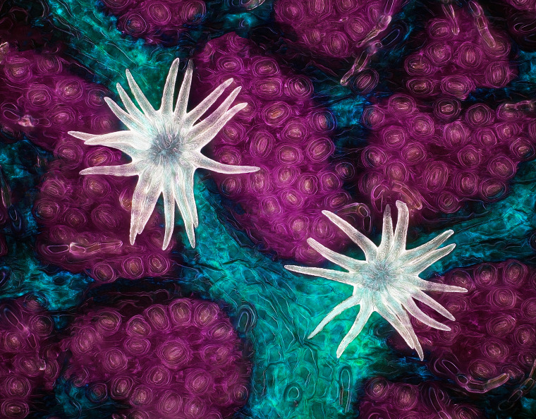
First place
Each year, the Nikon Small World photo contest fuses art and science to create mind-bending microscopic images.
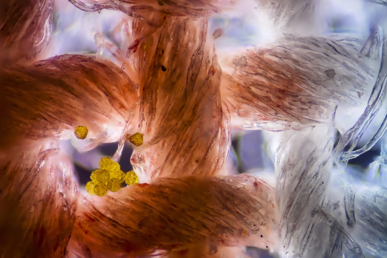
First place this year goes to Jason Kirk, of the Baylor College of Medicine, for his image of a southern live oak leaf's vessels, trichomes and stomata. The final image is the result of stacking 200 individual images to make the invisible visible.
The lighting proved to be especially tricky.
“Microscope objectives are small and have a very shallow depth of focus," Kirk said. "I couldn’t just stick a giant light next to the microscope and have the lighting be directional. It would be like trying to light the head of a pin with a light source that's the size of your head. Nearly impossible.”
He was able to make it work in post-production through adjusting the color temperature and hue.
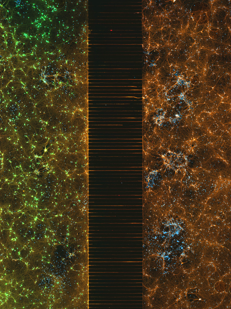
Second place
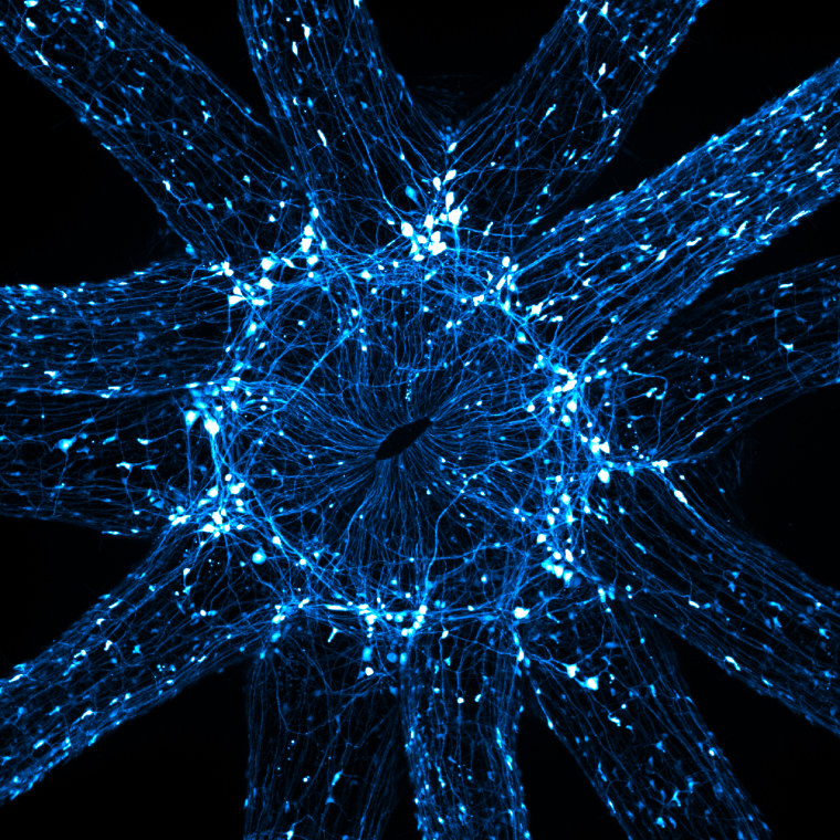
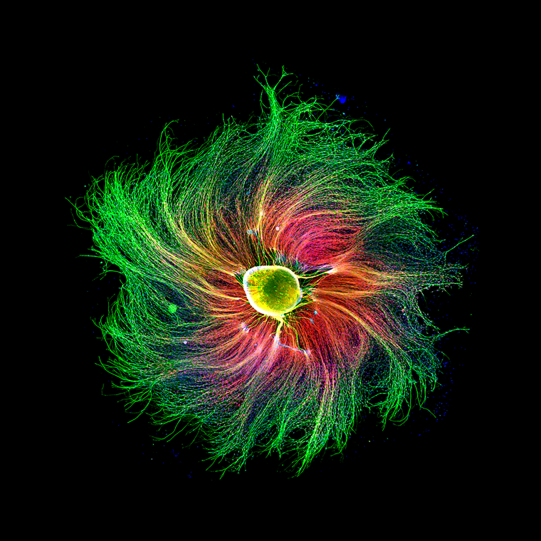
Second place was awarded to Esmeralda Paric and Holly Stefen of Macquarie University's Dementia Research Center for their image of a microfluidic device containing 300,000 networking neurons in two isolated populations. Both sides were treated with a unique virus and bridged by axons.
