Weird Science
Nikon photo contest reveals fantastic microscopic world that surrounds us
Tour psychedelic cellular landscapes, face off with a jumping spider, and more amazing views through the lens of a microscope.
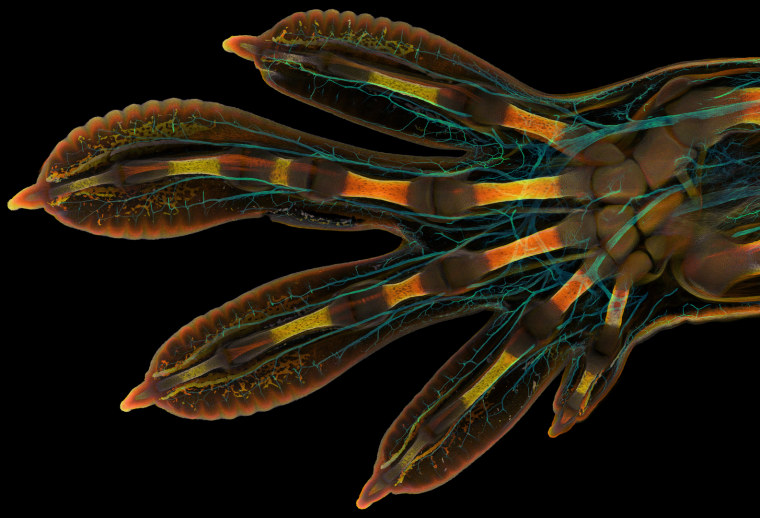
First place
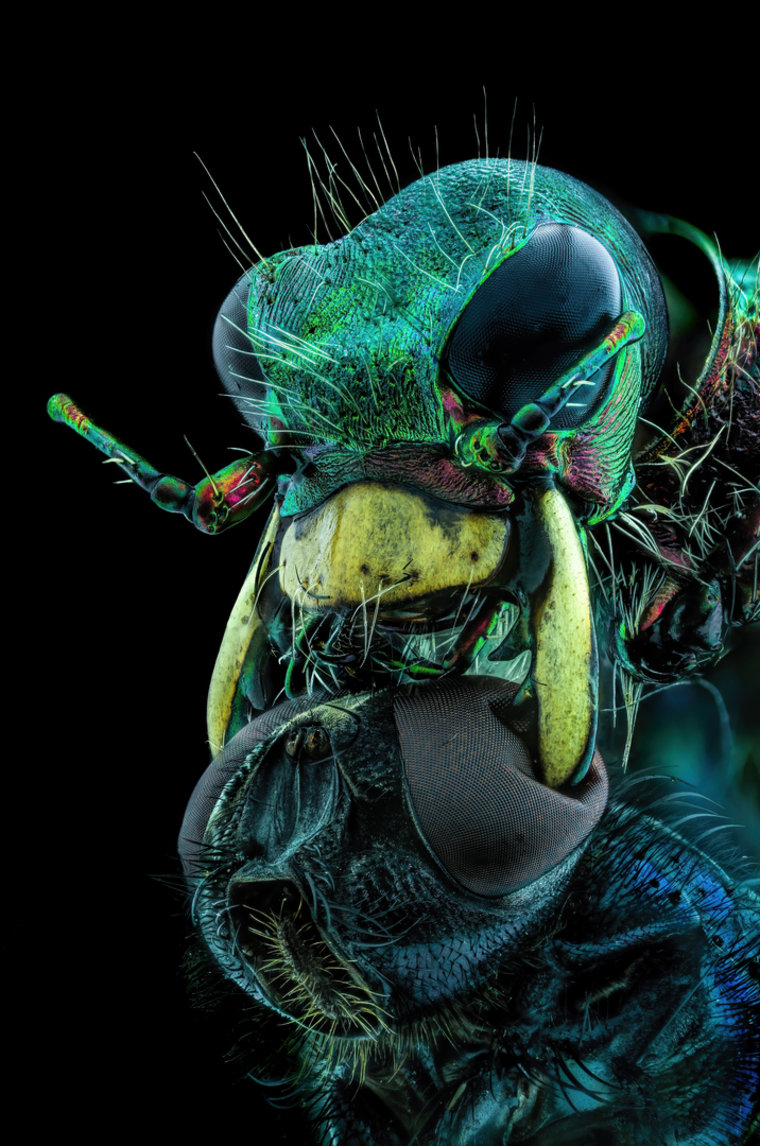
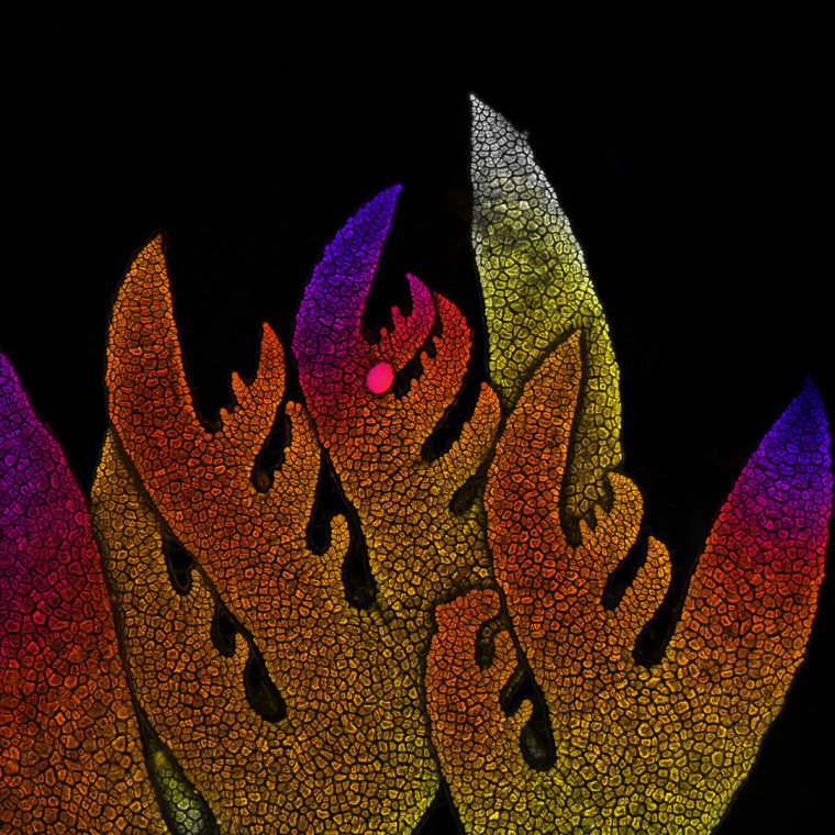
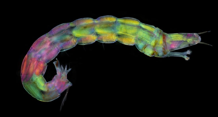
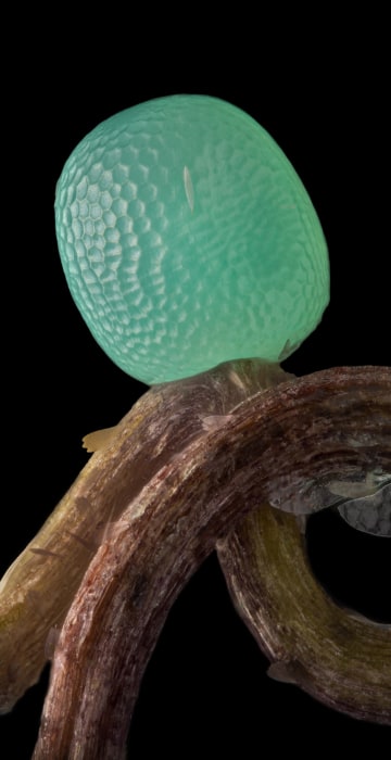
Each year, art and science collide in the Nikon Small World photo contest to produce breathtaking (and sometimes unnerving) microscopic images.
This year's first place prize was awarded to Grigorii Timin, supervised by Dr. Michel Milinkovitch at the University of Geneva, for his image of an embryonic hand of a Madagascar giant day gecko.
Timin used image stitching to merge hundreds of images together to create the final image. "The scan consists of 300 tiles, each containing about 250 optical sections, resulting in more than two days of acquisition and approximately 200 GB of data," said Timin.
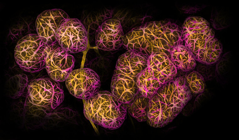
Second place
Second place was awarded to Dr. Caleb Dawson for his image of breast tissue showing contractile myoepithelial cells wrapped around milk-producing alveoli. Taking a week to process, the myoepithelial cells were stained with multiple rounds of fluorescent dyes and captured with a confocal microscope.
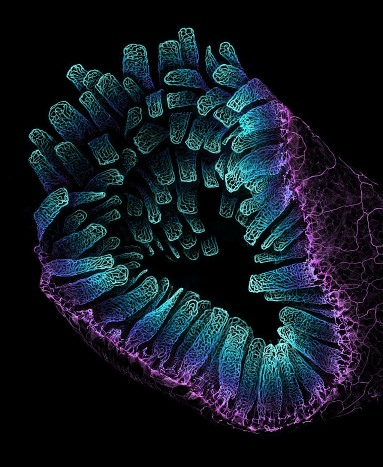
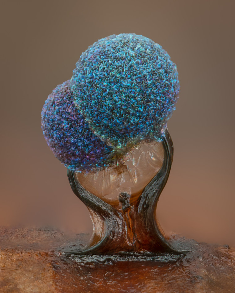
Slime mold
Lamproderma is a genus of slime mold.
Slime molds were once thought to be a kind of fungus, but later work revealed that these puddles of goo are part of a motley group of microbes known as protists.
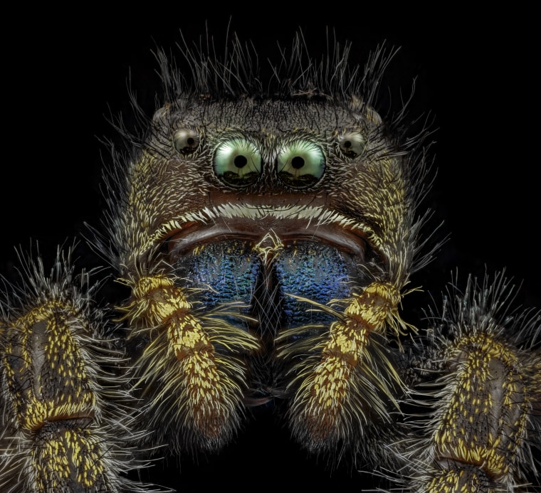
Bold jumping spider
Jumping spiders are a group of spiders that actively hunts its prey rather than trapping it in webs. Like most spiders, they have four pairs of eyes.
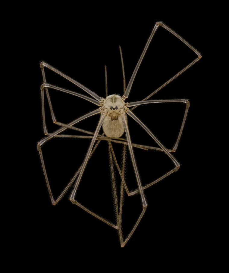
Daddy longlegs
Daddy longlegs (Pholcus phalangioides) have been skittering around the Earth for more than 300 million years.
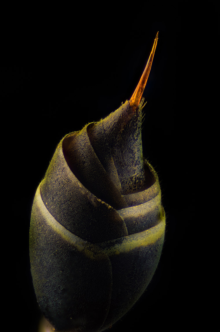
Wasp stinger
Stinger of a small paper wasp (Vespidae Protopolybia).
Unlike bees, wasps can sting multiple times because they don't leave their stinger behind.
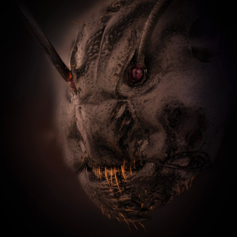
Face of an ant
Close-up of an ant (Camponotus).
